


In autoimmune research, regulatory T cells (Tregs) are generally regarded as the "brakes" that suppress excessive immune responses. However, a mouse study published in the Journal of Experimental Medicine shows that when Tregs lack the transcription factor Helios, the mice do not develop multi-organ inflammation; instead, they develop acquired lipodystrophy: adipose tissue is destroyed by autoreactive CD8⁺ T cells, which in turn leads to fatty liver and insulin resistance. The study further reveals that Helios, by directly regulating genes such as Id3, Tcf1, Lef1, and Bach2, restricts excessive differentiation of Tregs toward the effector stage and maintains their function after terminal differentiation. This is also a typical model of "increased Treg numbers but dysfunctional Tregs."
Research Background: Treg Dysfunction and Tissue Homeostasis
Tregs are a population of Foxp3-expressing CD4⁺ T cells responsible for maintaining immune tolerance and tissue homeostasis. In adipose tissue, the proportion of Tregs is higher than in lymphoid organs, and they possess a unique transcriptomic signature, participating in the control of metabolic balance.
Previous studies have found that in many autoimmune models, Treg numbers do not decrease but rather increase, yet their function is defective. Helios (encoded by Ikzf2) is a transcription factor in Tregs. Earlier research showed that mice with Treg-specific deletion of Helios develop systemic Th1 immune activation, reduced follicular regulatory T cells, and enhanced germinal centers.
However, a key question remains unanswered: after Helios deletion, at which step do Tregs actually "fail"? Why is the inflammation concentrated specifically in adipose tissue?
Research Objective: Tracing the Treg Differentiation Pathway After Helios Deletion
The goal of this study was to clarify the role of Helios in Treg effector differentiation, specifically:
First, why Helios-deficient mice develop lipodystrophy and metabolic disorders;
Second, what changes occur in the phenotype of Helios-deficient Tregs in adipose tissue;
Third, through which target genes Helios regulates Treg differentiation and function.
Research Methods (Brief)
The study used Treg-specific Helios knockout mice (Ikzf2fl/fl × Foxp3-Cre). Through metabolic cages, histology, flow cytometry, RNA sequencing, and ChIP sequencing, Tregs from wild-type and knockout mice were compared.
To verify the role of CD8⁺ T cells, the researchers crossed the knockout mice with β2-microglobulin-deficient mice to generate double-knockout mice lacking CD8⁺ T cells. To distinguish cell-intrinsic defects from the effects of the inflammatory environment, Foxp3 heterozygous female chimeric mice were also used.
Core Findings: Increased Numbers, Functional Failure
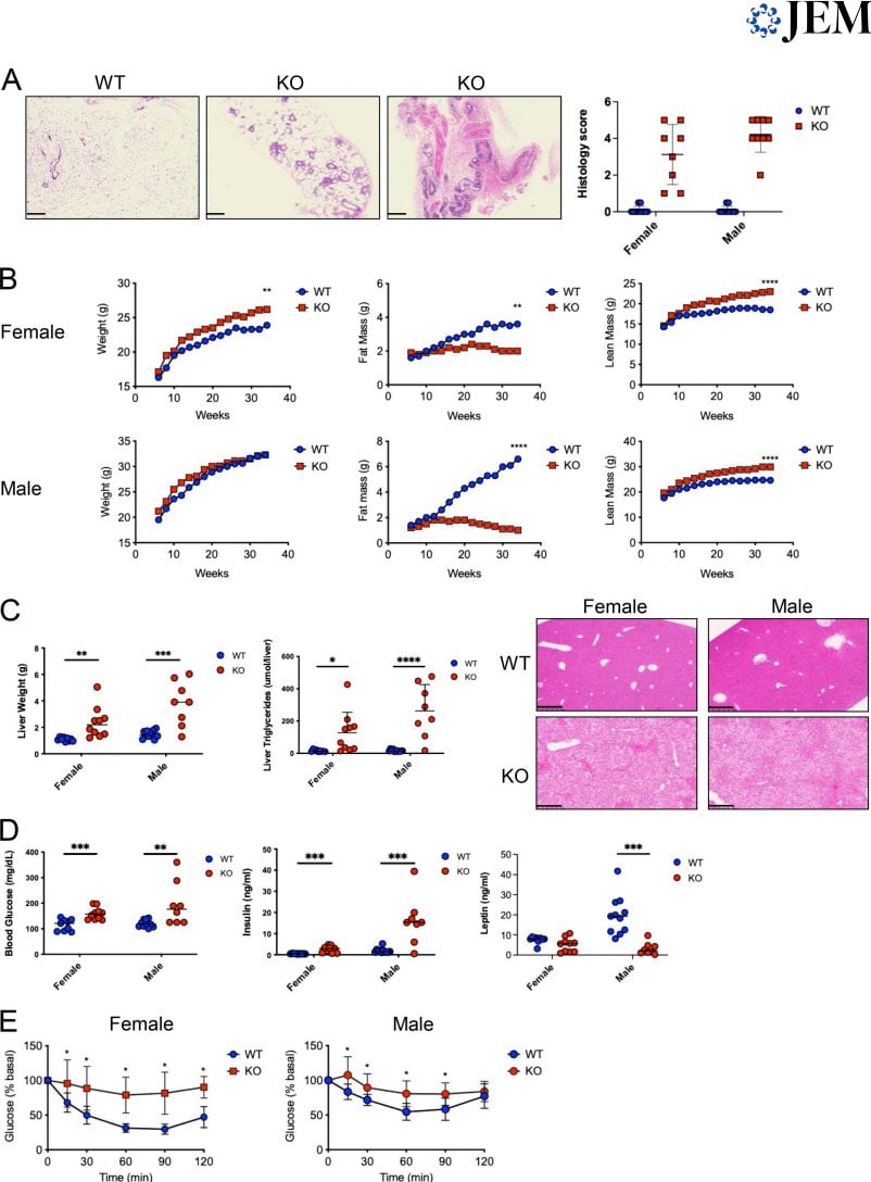
Adipose tissue is destroyed by CD8⁺ T cells. At 3 months of age, the adipose tissue of knockout mice showed massive infiltration of hematopoietic cells and eventually disappeared completely. CD8⁺ T cells in adipose tissue highly expressed CD107, granzyme B, IFNγ, and Fas-L, suggesting that they kill adipocytes through cytotoxic and Fas/Fas-L pathways. In double-knockout mice lacking CD8⁺ T cells, adipose tissue was preserved, and metabolic abnormalities were reversed.
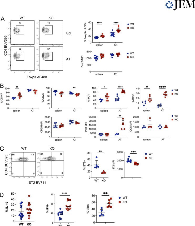
Abnormal adipose Treg phenotype. In the adipose tissue of knockout mice, the proportion of Tregs was elevated, but two terminally differentiated subsets, ST2⁺ and CXCR3⁺, were significantly reduced, and Klrg1 and IRF4 expression was also decreased. These Tregs displayed a CD62L⁻Tcf1⁻ effector phenotype, yet lacked terminal differentiation markers.
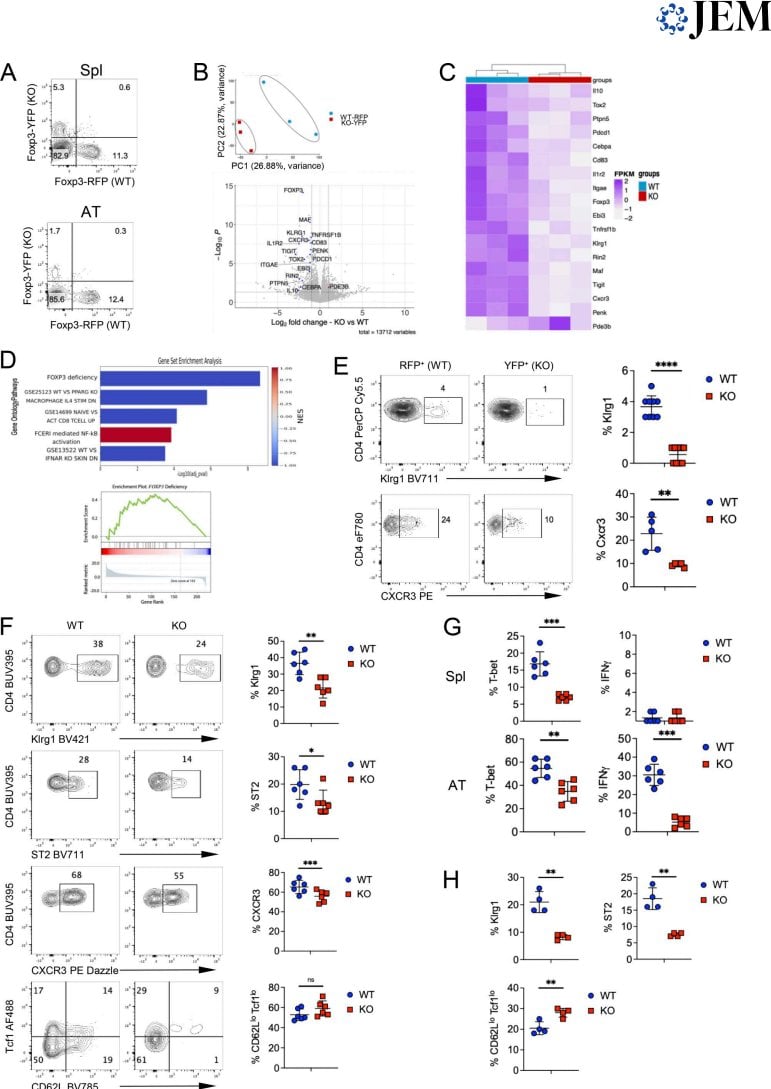
Helios directly regulates differentiation-restricting factors. ChIP-seq showed that Helios binds to the regulatory regions of Id3, Tcf7, Lef1, and Bach2. Together, these genes act to restrict Treg differentiation toward the effector stage. In Helios-deficient adipose Tregs, the expression of these genes was decreased, and effector differentiation thus went "out of control."
The defect is cell-intrinsic. In chimeric mice, even when wild-type Tregs were present to suppress inflammation, knockout Tregs still showed decreased expression of Klrg1, ST2, and CXCR3, indicating that this defect does not depend on the inflammatory environment.
Innovation or Breakthrough
The uniqueness of this study lies in its revelation of a paradoxical phenomenon: Helios deletion leads to increased Treg numbers and enhanced activation, yet dysfunctional Tregs.
Mechanistically, Helios does not simply suppress or promote Treg differentiation; rather, it plays two roles simultaneously: directly restricting effector differentiation while indirectly ensuring that terminal differentiation proceeds normally. After Helios deletion, Tregs "overshoot," entering the effector stage but failing to complete functional maturation.
In addition, this model directly links autoimmune lipodystrophy with CD8⁺ T cell-mediated killing of adipocytes for the first time, providing a new perspective for understanding tissue-specific autoimmunity.
Practical Implications: Autoimmunity Through the Lens of Treg Differentiation
This study suggests that Treg function cannot be assessed by numbers alone. When evaluating Treg-related diseases or therapeutic strategies, the "quality" of effector differentiation is equally critical.
Helios controls the pace of Treg differentiation through a core transcriptional network (Id3, Tcf1, Lef1, Bach2). If this network is disrupted, Tregs may lose their protective function in specific tissues.
For translational research, this model also reminds us that Tregs in different tissues depend on different transcriptional programs, and Tregs in adipose tissue especially depend on the terminally differentiated state maintained by Helios.
Summary and Reflections
The lipodystrophy model of Helios-deficient mice demonstrates that the suppressive function of Tregs requires precise regulation of differentiation. Increased numbers do not equal normal function, and excessive effector differentiation may instead lead to tissue damage.
This study also leaves an open question: in human autoimmune diseases, does a similar state of Treg "pseudo-activation" also exist? If so, can we identify those Treg subsets with normal numbers but impaired function by detecting the expression of specific transcription factors?