



Hereditary cerebellar ataxias have long been regarded as a group of neurodegenerative diseases that are difficult to intervene in. When the causative gene encodes a secreted synaptic organizing factor, the situation may be entirely different. Recent research reveals that in a class of early-onset ataxia caused by biallelic mutations in the CBLN1 gene, the core mechanism is unexpectedly a scarcity of an extrasynaptic factor—and this state can be reversed in adulthood through AAV-mediated gene delivery.
From Clinical Phenotype to Causative Gene: Key Clues from Two Families
The research team identified patients with early-onset cerebellar ataxia in two consanguineous Egyptian families. Symptoms first appeared at 3-4 months of age, presenting as abnormal eye movements and motor developmental delay. As the disease progressed, the affected children developed truncal and limb ataxia, hypotonia, and progressive gait impairment. Brain MRI showed cerebellar atrophy, most prominent in the vermis.
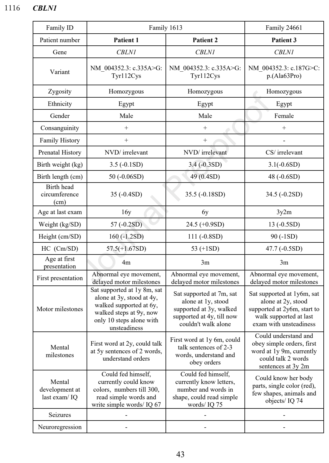
Whole-genome sequencing identified homozygous missense mutations in the CBLN1 gene in the two families: c.187G>C (p.A63P) and c.335A>G (p.Y112C). Sanger sequencing confirmed co-segregation of the mutations with the disease, consistent with an autosomal recessive inheritance pattern.
The Cbln1 protein encoded by CBLN1 is a key organizing factor of the parallel fiber–Purkinje cell (PF-PC) synapse. As a secreted glycoprotein, it bridges the presynaptic neurexin and the postsynaptic glutamate receptor δ2 (GluD2), and is essential for synapse formation and long-term maintenance. Previously, Cbln1 knockout mice exhibited severe PF-PC synaptic defects, but the gene had never been confirmed to be associated with human disease.
Mutations Cause a Functional Extracellular Deficiency
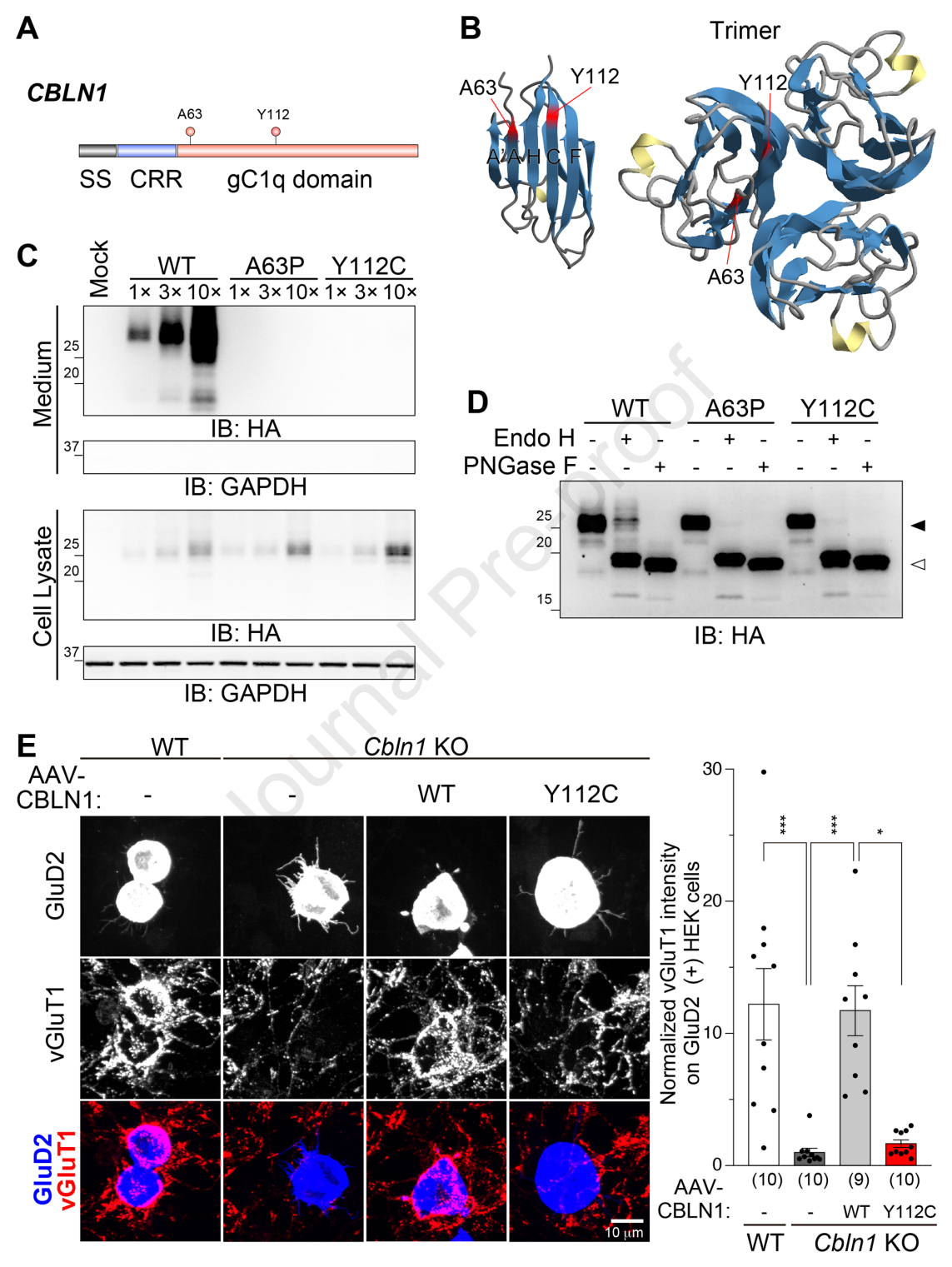
Structural analysis localized the two residues A63 and Y112 near the subunit contact interface of the CBLN1 trimer. This suggests that the mutations may disrupt protein oligomerization, thereby affecting stability, intracellular trafficking, and secretion. In vitro experiments confirmed this hypothesis: the steady-state protein levels of both mutants in HEK293 cells were significantly reduced, and secreted protein was barely detectable in conditioned medium.
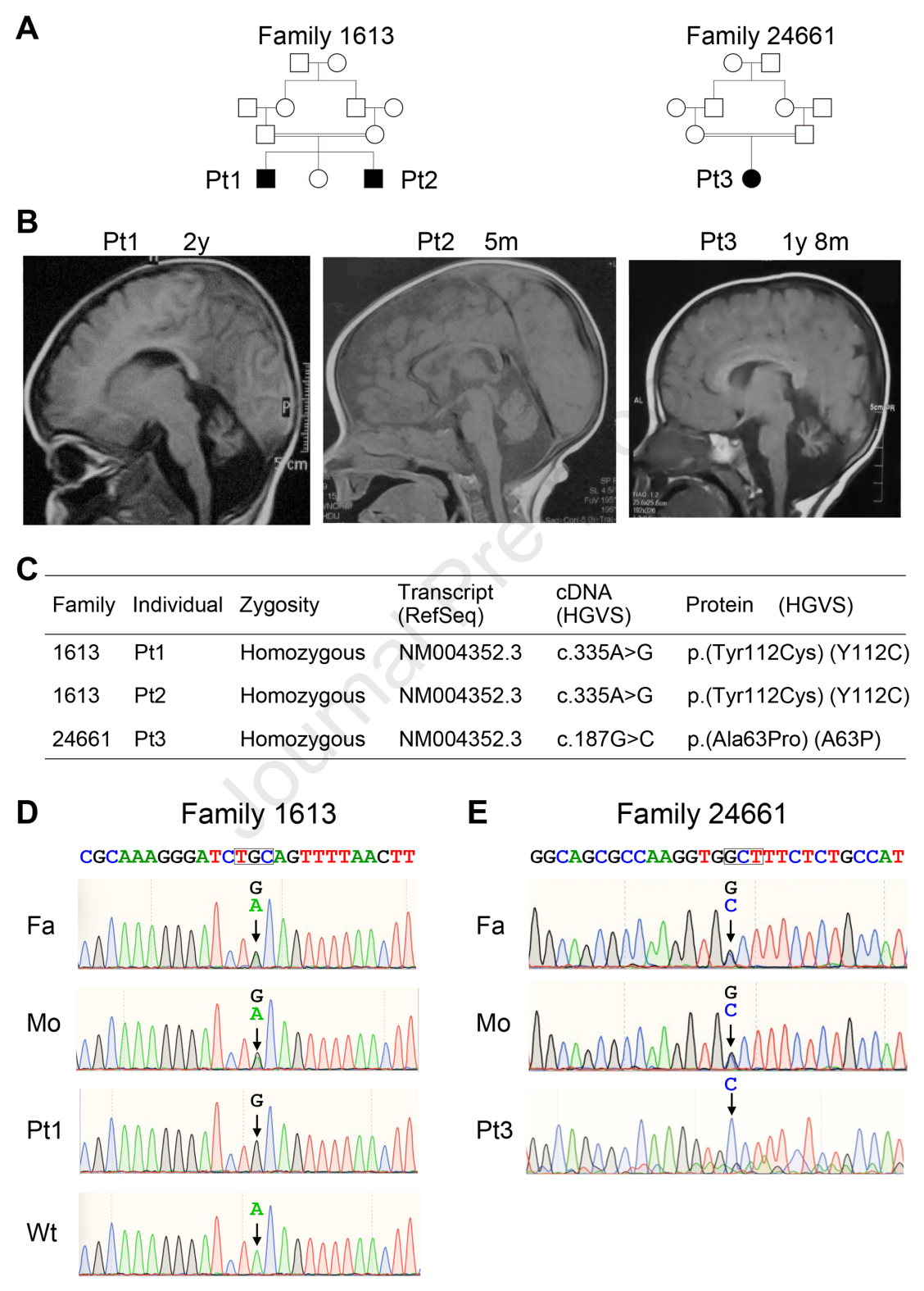
Glycosylation analysis revealed a more refined mechanism. Wild-type CBLN1 contains a mature glycoform resistant to endoglycosidase H, indicating that the protein has completed processing through the endoplasmic reticulum/cis-Golgi. In contrast, the mature glycoform of both mutants was significantly reduced, indicating that the protein is retained in the early secretory pathway. This anterograde trafficking defect directly leads to a substantial decrease in extracellular CBLN1 availability.
Functional experiments further validated this conclusion. In an artificial synapse formation assay, cerebellar granule cells expressing CBLN1-Y112C failed to induce presynaptic differentiation on cells expressing GluD2, consistent with the behavior of Cbln1 knockout cells. This indicates that the mutant protein is functionally equivalent to a complete deficiency.
Knock-in Mouse Model: From Molecular Defects to Behavioral Phenotypes
To validate the disease mechanism in vivo, the researchers generated Cbln1 knock-in mice carrying the Y112C mutation. Homozygous mutant mice were born at Mendelian ratios, and the cerebellar lobular structure and laminar organization were largely preserved, but cerebellar volume was significantly reduced—the vermis cross-sectional area was decreased by approximately 28% compared with wild type, consistent with the patients' imaging findings.
Immunohistochemistry showed that synaptically localized Cbln1 signal was barely detectable in the cerebellar cortex of knock-in mice, highly similar to Cbln1 knockout tissue. Behavioral analysis revealed a series of deficits: mutant mice fell from the rotarod within seconds, gait analysis showed abnormal hindlimb coordination, and video oculography recorded abnormally increased spontaneous eye movements—precisely recapitulating the patients' motor and cerebellar oculomotor phenotypes.
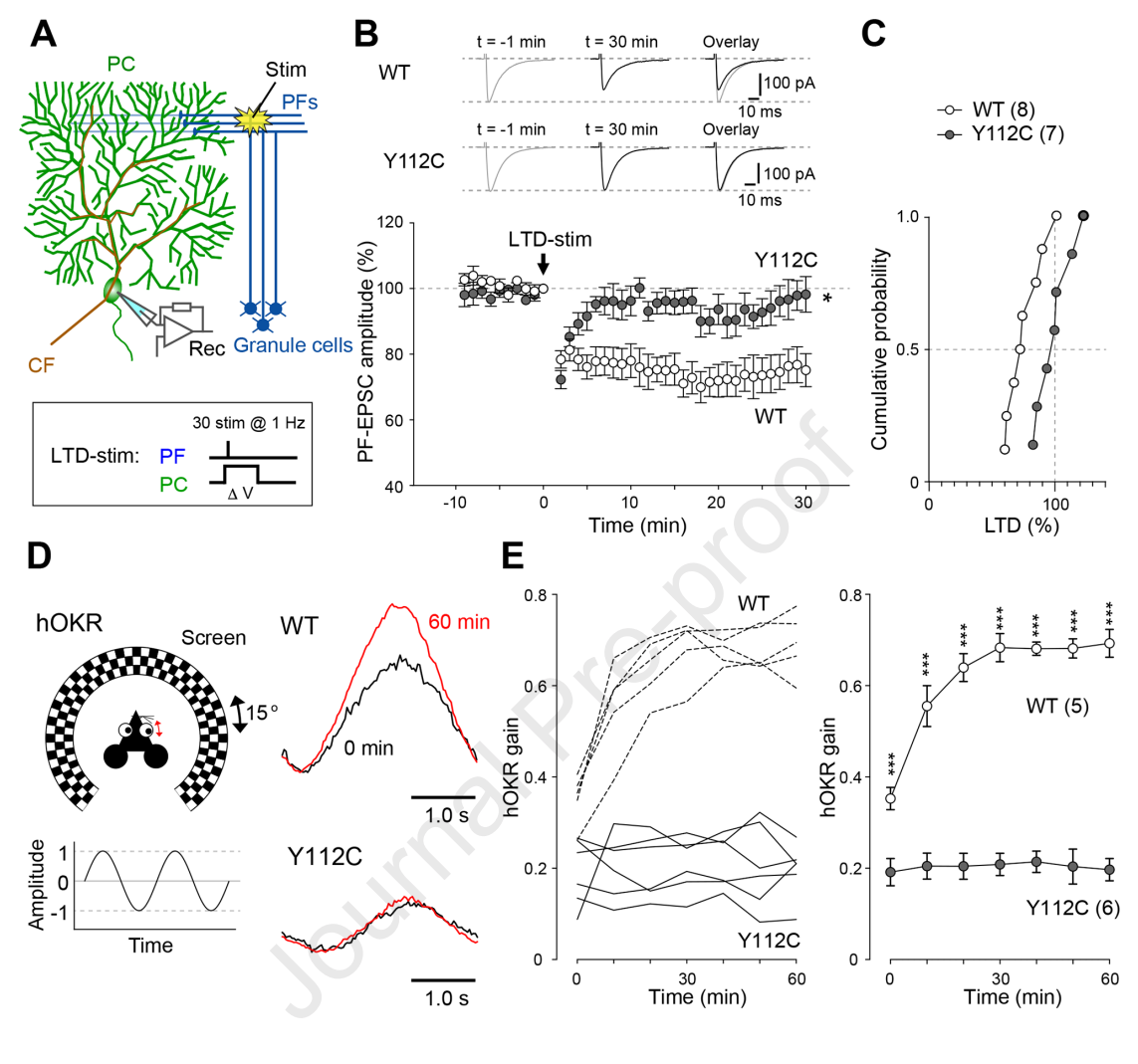
Electrophysiological recordings further defined the synaptic-level abnormalities. The amplitude of PF-evoked EPSCs in mutant mice was significantly reduced, and paired-pulse facilitation was enhanced, suggesting a decreased presynaptic release probability. More critically, PF-PC long-term depression (LTD), a core mechanism of cerebellar motor learning, was completely absent in mutant mice, and optokinetic reflex (OKR) adaptation was also severely impaired.
Astrocyte-Targeted AAV: A Reversible Strategy for Repair in Adulthood
Direct delivery of recombinant Cbln1 protein transiently improved motor function in mutant mice, but the effect lasted only about one week. This prompted the researchers to explore strategies capable of providing sustained extracellular CBLN1. A key design decision was the choice of cell source: attempts to directly drive CBLN1 expression in granule cells failed due to insufficient promoter activity, so the researchers turned their attention to astrocytes.
Astrocytes are in close proximity to PF-PC synapses in the molecular layer, and Bergmann glial processes wrap around synaptic structures, constituting an ideal local secretory microenvironment. More importantly, CBLN1 acts extracellularly, and its efficacy depends primarily on extracellular availability and synaptic targeting, rather than necessarily being expressed from an endogenous neuronal source. Restricting the transgene to non-neuronal cells may also reduce the risk of ectopic neuronal expression interfering with synapse formation.
The researchers constructed an AAV vector in which HA-CBLN1 expression is driven by the astrocyte-specific promoter GfaABC1D, and delivered it systemically to adult mutant mice via retro-orbital injection. Immunohistochemistry confirmed widespread cerebellar transduction, with HA signal enriched in the molecular layer and co-localized with GluD2, indicating that the secreted CBLN1 successfully reached synaptic sites.
The therapeutic effects were striking. In treated mice, PF-PC pre- and postsynaptic alignment improved, PF transmission function was restored, CF monoinnervation was re-established, and LTD was successfully re-induced. At the behavioral level, mutant mice recovered OKR adaptation, rotarod and treadmill performance approached wild-type levels, and abnormal spontaneous eye movements were corrected. These improvements were quantitatively confirmed 1-2 months after injection, with some phenotypic improvements persisting for at least 6 months.
Process Implications and Open Questions
The value of this study lies not only in establishing CBLN1 deficiency as a treatable synaptopathy, but also in providing multiple implications for AAV process development.
Strategic significance of target cell selection: When a therapeutic protein functions extracellularly, the endogenous expressing cell is not the only option. Astrocytes, as an alternative production source, offer the advantages of both anatomical proximity and widespread distribution. This suggests that process development should systematically evaluate the suitability of different promoter-cell combinations, rather than being limited to pursuing cell-type specificity.
Delivery route and biodistribution: Systemic delivery achieved widespread cerebellar transduction, but also produced ectopic expression in extracerebellar regions such as the hippocampus and cerebral cortex. This distribution differs from the endogenous Cbln1 pattern and may bring unknown extracerebellar effects. For process development, this means that a more refined vector biodistribution assessment system needs to be established to distinguish therapeutic distribution from potentially risky distribution.
Persistence trade-off between protein replacement vs gene delivery: Direct delivery of recombinant protein has transient effects, whereas AAV-mediated sustained expression offers the possibility of long-term repair. This contrast reinforces the process value of gene delivery in scenarios requiring sustained secreted protein, and also suggests that half-life and dosing frequency should be core parameters in the selection of delivery strategies.
Mechanistic significance of adult-stage reversibility: The extensive phenotypic reversal indicates that a considerable portion of the pathological changes are functional rather than structural damage. This has direct implications for patient selection and clinical trial design—even when imaging has already shown cerebellar atrophy, functional repair at the synaptic level may still confer clinical benefit.
A question worth pondering: does the success of astrocytes as a production source for secreted proteins mean that other neurological diseases caused by defects in secreted synaptic organizing factors could also adopt a similar strategy? When the therapeutic target shifts from within neurons to the synaptic cleft, do our requirements for vector tropism and expression persistence need to be redefined?