


A phase 1 trial in patients aged 12–18 years with severe hemophilia B has yielded an intriguing signal: factor IX activity peaks higher than in adults, but declines more markedly by week 52. This "high peak, steep slope" difference brings the issue of immune management in AAV gene therapy to the forefront.
Study Background and Design
Hemophilia B is caused by variants in the F9 gene; patients experience recurrent spontaneous bleeding, especially in joints and muscles, progressively developing arthropathy. Although regular prophylactic infusion can improve outcomes, lifelong injections and the cost burden still affect quality of life.
More critically, once arthropathy has developed, it may be irreversible even with regular treatment. Therefore, early intervention has become a consensus direction in the field.
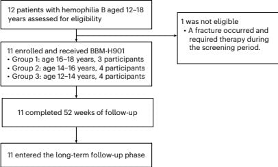
The three approved hemophilia B gene therapy products all cover only adults, and safety and efficacy data in the adolescent population remain lacking. This study enrolled 11 male adolescents aged 12–18 years, all with severe or moderately severe disease (FIX:C ≤ 2 IU dl⁻¹).
All subjects received a single intravenous infusion at a dose of 5 × 10¹² vg kg⁻¹, with 52 weeks of follow-up. The primary endpoint was safety.
Safety: No Dose-Limiting Toxicity
No dose-limiting toxicity was observed in the study. A total of 92 adverse events were reported within 52 weeks; the most common were elevated white blood cells (63.6%), elevated neutrophils (45.5%), and rash (27.3%), all associated with glucocorticoid use.
One serious adverse event was acute exacerbation of chronic apical periodontitis, unrelated to treatment, during which FIX:C remained stable. One subject developed elevated transaminases (ALT 197.0 U l⁻¹), which returned to normal within 4 weeks after immunosuppressive treatment, but FIX:C rose only transiently and fell to 6.2 IU dl⁻¹ at week 52.
All subjects developed high-titer AAV capsid-binding antibodies and neutralizing antibodies after treatment, persisting throughout the follow-up period.
Efficacy: Higher Peak, Faster Decline
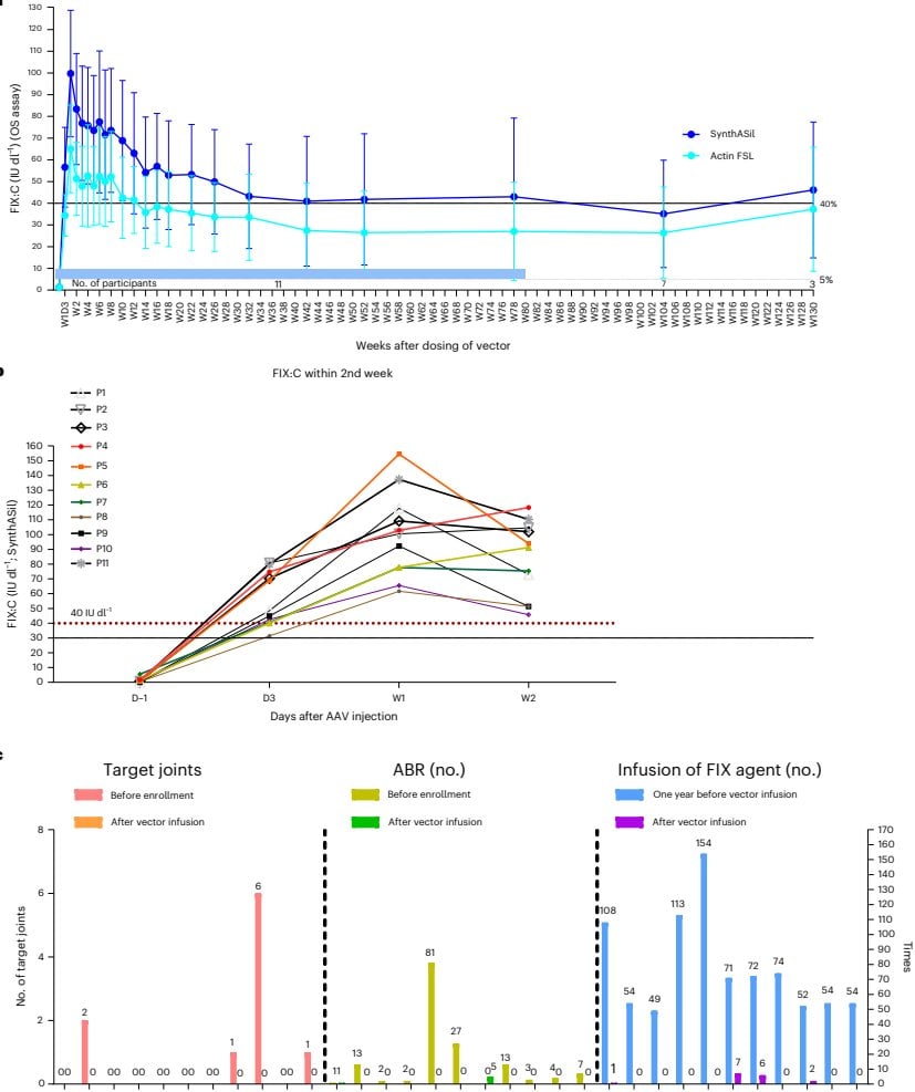
FIX:C rose rapidly after infusion: reaching 51.2 IU dl⁻¹ on day 3, peaking at 92.8 IU dl⁻¹ on day 6, and reaching 41.8 IU dl⁻¹ at week 52.
The annualized bleeding rate decreased from 13.9 before treatment to 0.5. 81.8% of subjects achieved zero bleeding, whereas at baseline this proportion was only 9.1%. The annual number of FIX infusions decreased from 71 to 0.
Compared with adult phase 3 data, adolescents had a higher peak (106.4 vs 97.7 IU dl⁻¹), but a more pronounced decline at week 52 (41.8 vs 55.1 IU dl⁻¹). The study suggests that adolescent hepatocytes proliferate actively, and AAV exists in the form of free episomes, which may be diluted by cell division—this hypothesis remains to be verified.
Notably, 72.7% of adolescents showed a marked decline at 2–4 weeks, whereas adults mostly showed a single decline only at 8–14 weeks. Whether this reflects an immune-mediated early effect currently remains undetermined.
Immune Mechanism: Steroid Withdrawal Is a Hurdle
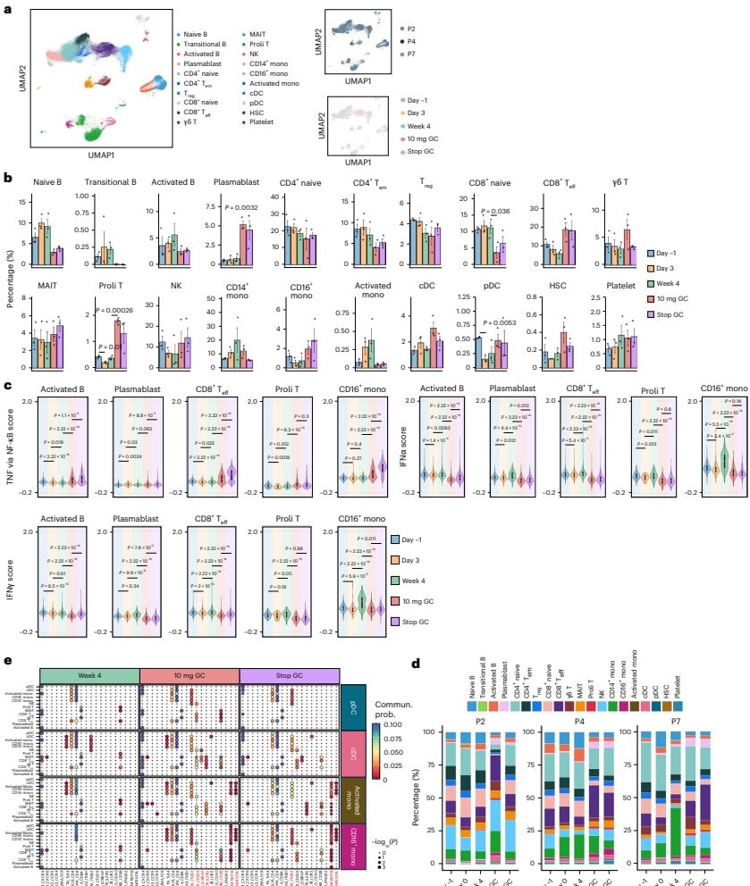
The study performed single-cell sequencing on 3 subjects, tracking immune changes at 5 time points: before infusion, 3 days after infusion, 4 weeks, when steroids were tapered to 10 mg, and when steroids were completely discontinued.
The results showed clear phases: when steroids were tapered to 10 mg, the FOS gene was highly expressed, the immune system was in a state of antigen presentation and NF-κB priming, and T cell activation was relatively mild; after complete discontinuation of steroids, the interferon and NF-κB pathways were synergistically activated, with significant T cell activation.
Both subjects with persistently declining efficacy exhibited this steroid withdrawal-induced T cell activation. The study suggests that during the steroid tapering phase, a combined immunosuppressive regimen may be necessary.
Implications for Process and Manufacturing
This study raises several questions worth considering for AAV vector process development.
First, vector persistence in rapidly proliferating tissues. Adolescent hepatocytes divide actively; if the episome dilution hypothesis holds, then long-term expression of the vector depends not only on transduction efficiency but also on the proliferation dynamics of target cells. This has implications for dose selection and vector design.
Second, the relationship between empty capsid rate and immunogenicity. The study did not directly compare empty capsid proportion with immune response, but high-titer antibodies appeared in all subjects, suggesting that management of the immunogenicity of the capsid itself remains a core issue.
Third, batch consistency and clinical comparability. Could the efficacy difference between adolescents and adults partly stem from differences in vector batches or assay methods? In the study, FIX:C was measured simultaneously with two reagents, SynthASil and Actin FSL, with a ratio of 1.49, indicating that the assay system itself introduces significant bias.
Fourth, the impact of purification processes on the immune profile. Residual host cell proteins, nucleic acids, or empty capsids from different purification routes may shape different immune microenvironments. This may be even more worth attention in populations with more active immune systems, such as adolescents.
Is the "high peak, steep slope" of adolescent gene therapy due to physical dilution from hepatocyte proliferation, early mobilization of the immune system, or a combination of both? The answer to this question may determine the direction in which next-generation AAV vector process optimization should proceed.